Recent News
NQVL Design Phase Award
October 1, 2025
CHTM Joins NSF's NQVL Pilot Projects
August 9, 2024
OSE PHD, Dr. Xuefeng Li - Wins The Outstanding Interdisciplinary Graduate Programs Award
May 10, 2024
Dr. Ali Rastegari - 2024 OSE Best Dissertation Award Winner
May 10, 2024
News Archives
UNM professor developing new, super-resolution light-sheet microscopy technique
November 7, 2016 - Excerpted from an article by Aaron Hilf in the UNM Newsroom

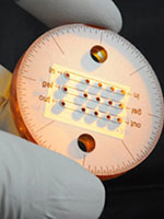
The Lidke group worked with Sandia National Labs to develop microfluidics chips that contain an integrated mirror to allow their light-sheet technique to work.
UNM associate professor of Physics & Astronomy and Optical Science & Engineering faculty member Keith Lidke has developed a new technique, single objective light-sheet microscopy, that improves on an existing method of flourescence microscopy. His findings were published in Biomedical Optics Express this year.
For scientists developing life-saving medicines, knowing how cells interact and communicate with one another is important. Being able to see those interactions through a microscope hasn’t always been possible. A new approach to this technique, developed by Lidke, his research group and collaborators at Sandia National Laboratories, allows researchers a better view of cellular interactions. The technique has the ability to help answer questions about how cells communicate and work internally, making it possible for researchers to develop medicine or therapies that utilize these interactions.

Keith Lidke, UNM Physics & Astronomy associate professor
According to Lidke, traditional fluorescence microscopy techniques can only provide researchers a very limited view of the cells they are viewing and exposes the sample to an abundance of light that degrades the image quality and leads to cell damage through photo-toxicity. The new approach overcomes these prominent issues and allows light-sheet microscopy to be performed using common microscopes found in most cell biology laboratories.
“What light-sheet microscopy does is allow us create a sheet of light that is matched exactly to the focal plane that we’re imaging,” explained Lidke. “We reduce light exposure and we reduce background noise in the system, so in living cells that allows us to see fluorescent proteins with enough signal to look at the dynamics of those proteins.”
While Lidke’s technique is still in its early stages, he has already received a lot of interest from researchers at UNM and across the country because of the unique view the equipment can provide.
“Knowing that our work has a potentially valuable application really makes what we’re doing everyday feel extremely important,” Lidke said.

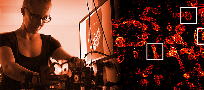
Post-doc Marjolein Meddens works with Lidke research group on light-sheet microscopy.
Read the original article to learn how this technique gives researchers the opportunity to see cellular interaction on an entirely new level:
Physics professor developing super-resolution microscopy techniques, by Aaron Hilf, UNM Newsroom


