

Recent News
CHTM Joins NSF's NQVL Pilot Projects
August 9, 2024
OSE PHD, Dr. Xuefeng Li - Wins The Outstanding Interdisciplinary Graduate Programs Award
May 10, 2024
Dr. Ali Rastegari - 2024 OSE Best Dissertation Award Winner
May 10, 2024
2024 OSE Spring and Summer Graduates
May 10, 2024
News Archives
Nanoscale physics research team performs magnetic resonance spectroscopy on a diamond chip
September 30, 2016 - Sharon Steely for CHTM
Victor Acosta
A grant from the National Science Foundation (NSF) began on January 1, 2016, for research conducted by the Quantum Nanophotonic and Biosensing Team at the Center for High Technology Materials (CHTM) at The University of New Mexico (UNM). The grant was awarded through Small Business Technology Transfer (STTR), an NSF program whose most important role is to bridge the gap between performance of basic science and commercialization of resulting innovations.
The STTR Phase I grant award of $225,000 for the research is shared with the Principal Investigator (PI) for the project, Andrejs Jarmola of ODMR Technologies, Inc., which is a Berkeley-based start-up developing chip-scale diamond platforms for trace magnetic resonance spectroscopic analysis. The CHTM research team is led by Victor Acosta (Co-PI), UNM Physics & Astronomy assistant professor and CHTM faculty member. A unique feature of the STTR program is the requirement for the small business to formally collaborate with a research institution in Phase I and Phase II. The CHTM team conducts basic applied physics research in collaboration with its industrial partner.
Sensor to provide chemical detection with trace samples
The project is to produce an optically-detected magnetic resonance sensor for determining the chemical composition of trace quantities of liquid and powder samples. This is critical in numerous industries ranging from defense forensics and environmental safety, to materials synthesis and petroleum exploration. This new sensor will have a direct and broad impact on public security and defense, for example in airport and border security checks. The reliable detection and analysis of small quantities of potentially-threatening chemicals and materials are essential for simultaneously meeting the increased demands for security and eliminating the need to test large quantities of analyte (the substance being measured). An impact on science education is also anticipated, based on the anticipated development of an affordable turnkey system, which can be used in instructional and research settings. 2
Detection of biomolecules at 10,000 times greater sensitivity than current devices
The research is also funded by the National Institutes of Health (NIH) in the amount of $216,281. The nanophotonic magnetic resonance spectroscopy platform is capable of uniquely detecting minute quantities of biomolecules. The specific detection of biomolecules plays a central role in modern life science including cell systems biology, high throughput drug screening, and clinical oncology. In addition to fluorescence microscopy, nuclear magnetic resonance, and radiation techniques, electron paramagnetic resonance (EPR) has emerged as a nearly background-free molecular sensing modality. The goal of the proposed research is to develop a new type of sensor to detect the type and behavior of biomolecules, such as proteins, with 10,000 times better sensitivity than current devices. The benchtop system is anticipated to be used for high throughput analysis of proteins in cell systems biology. In the longer term, the technology may be used in a miniature ultra sensitive sensor for malaria diagnosis. 3
Quantum Sensing
Three aspects of quantum sensing will produce a dramatic improvement over traditional EPR, which detects the small net magnetization of electron spins: 3
- Detection of nanoscale variations in magnetization will produce orders of magnitude larger signals at ambient temperature.
- A magneto optical diamond film is used to transduce the EPR signal into the optical domain.
- Gratings are nanofabricated on the diamond surface to enhance contact between the sensor and analyte by more than >10x. 3
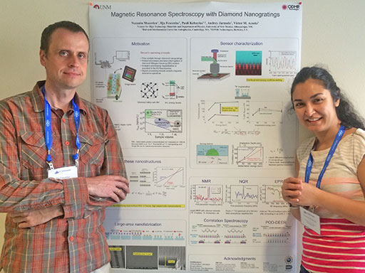
Postdoc Ilja Fescenko (l) and graduate student Nazanin Mosavian (r) with the poster they presented at the Rocky Mountain Magnetic Resonance Conference in July. Nazanin won the Jack E. Crow Travel Grant to present an invited talk at the joint session to over 200 scientists. Ilja won the postdoctoral travel award to present his own invited talk in front of the same audience.
Taking the research to market
UNM's Science and Technology Corporation (STC.UNM) is responsible for transferring technologies developed at UNM to the marketplace. STC has filed for intellectual property and is exploring commercialization options for the product of the research, a magnetic-resonance imaging spectrometer which detects sub-nanoliter volumes of liquid, gas, and thin-film samples over a wide range of ambient temperatures. 4
The sensor is structured to increase the total contact area with the sample, causing a boost in the Nanophotonic Magnetic Resonance (NMR) signal and reduction in acquisition time. This allows for sensitive detection of sub-nanoliter volumes of trace samples under a wide variety of environmental conditions. In addition, this technique is capable of being employed in low magnetic fields to detect nuclear quadrupole resonance (NQR), making it possible to detect extremely small trace quantities of analytes (e.g. nanograms of powder). 4
First setup of its kind at CHTM: Nanophotonic Magnetic Resonance (NMR) Spectroscopy
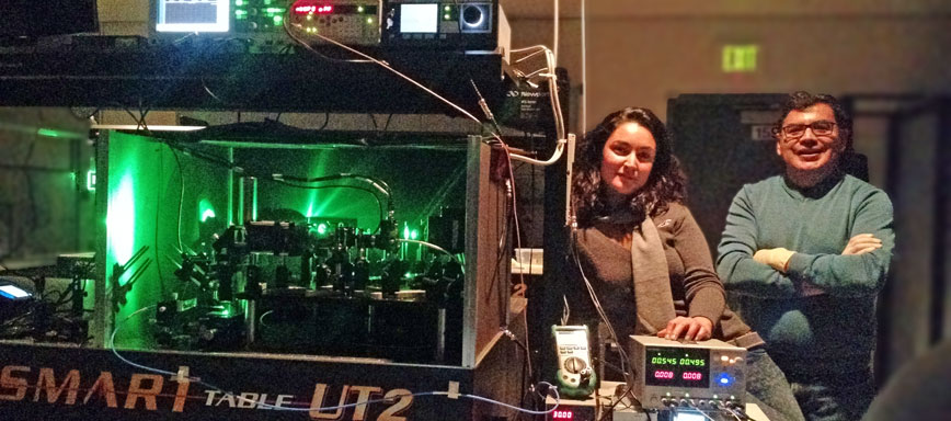
Graduate student Nazanin Mosavian (l) and postdoc Francisco Benito (r) (now an engineer with Intel) with the first NMR Spectroscopy setup at CHTM. There are now three NMR Spectroscopy setups at CHTM.
The research: Diamond Nanophotonics
Republished with permission from the Quantum Nanophotonics and Biosensing research team's website.
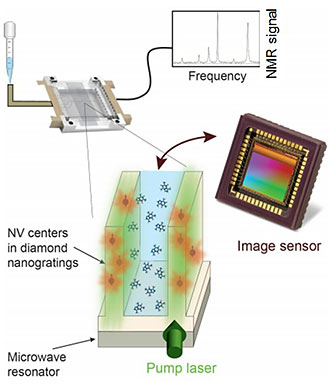
Schematic of the microfluidic spectrometer
1. Nanophotonic magnetic resonance spectroscopy
This project is led by UNM graduate student Nazanin Mosavian, Postdoc Ilja Fescenko, and Research Assistant Professor Abdelghani Laraoui in collaboration with ODMR Tech CEO Andrey Jarmola, Harvard scientist Pauli Kehayias, JGU-Mainz researchers Dmitry Budker and Lykourgos Bougas, and UNM scientists Steve Brueck and Alexander Neumann.
In this project, we apply the basic physics of color centers in diamond to create a device with unique properties desirable for chemical trace analysis. Unlike many magnetic-resonance (MR) systems, this spectrometer operates at ambient temperature, in a range of magnetic fields easily generated by small permanent magnets, and with nL sample volumes.
The analyte is delivered via a microfluidic chip to a sensor region consisting of a nanostructured diamond doped with nitrogen-vacancy (NV) color centers [1-3]. By applying pulses of laser light and microwaves, the magnetic field from the analyte’s precessing magnetization becomes encoded in the NV fluorescence signal. Analysis of the fluorescence signal reveals the analyte's MR spectrum, from which the chemical composition can be extracted from established libraries.
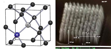
Diamond NV center and nanophotonic structures
1. V. M. Acosta, D. Budker, P. R. Hemmer, J. R. Maze, and R. L. Walsworth, "Optical magnetometry with nitrogen-vacancy centers in diamond", Cambridge University Press (2013).
2. V. M. Acosta, E. Bauch, M. P. Ledbetter, C. Santori, K. M. C. Fu, P. E. Barclay, R. G. Beausoleil, H. Linget, J. F. Roch, F. Treussart, S. Chemerisov, W. Gawlik, D. Budker, “Diamonds with a high density of nitrogen-vacancy centers for magnetometry applications.” Physical Review B 80, 115202 (2009).
3. A. Jarmola, V. M. Acosta, K. Jensen, S. Chemerisov, D. Budker, “Temperature and magnetic field dependent longitudinal spin relaxation in nitrogen-vacancy ensembles in diamond.” Physical Review Letters 108, 197601 (2012).
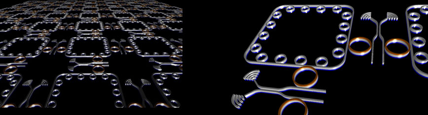
2. Few-photon optical logic in scalable networks
This project is currently led by UNM graduate students Yaser Silani and Forrest Hubert.
A naive extrapolation of Moore’s law predicts that computational elements will reach the 1 nm regime (~4 atoms across) by 2030. Further improvements will require new paradigms for information processing. One approach is to use all-optical logic gates to process information encoded in the amplitude and phase of light. This approach differs from previous failed attempts by using integrated nanophotonic networks to perform logic with the lowest power consumption allowed by quantum mechanics — the single photon level.
All-optical logic requires that photons interact. This is a challenge, since photons lack charge, mass, etc., but the photon-photon interaction can be mediated by a third party, atoms. In our case, all-optical logical operations will be performed by nonlinear optical addressing of color centers in diamond that are coupled to nanophotonic cavities [1-3]. Sequential logic will be enabled by embedding these cavities into interacting networks of optical waveguides, beamsplitters, and other on-chip photonic elements. An artist's rendering is depicted below, courtesy of C. Santori, HP Labs.
1. V. M. Acosta, P. R. Hemmer, “Nitrogen-vacancy centers: Physics and applications.” MRS Bulletin 38,127 (2013).
2. V. M. Acosta, K. Jensen, C. Santori, D. Budker, R. G. Beausoleil, “Electromagnetically-induced transparency in a diamond spin ensemble enables all-optical electromagnetic field sensing.” Physical Review Letters 110, 213605 (2013).
3. A. Faraon, C. Santori, Z. Huang, V. M. Acosta, R. G. Beausoleil, “Coupling of Nitrogen-Vacancy Centers to Photonic Crystal Cavities in Monocrystalline Diamond.” Physical Review Letters 109, 033604 (2012).
4. A. Faraon, C. Santori, Z. Huang, K.-M. C. Fu, V. M. Acosta, D. Fattal, R. G. Beausoleil, “Quantum photonic devices in single-crystal diamond.” New Journal of Physics 15, 025010 (2013).
3. Nanodiamond molecular imaging
4. Bio-magnetic imaging
Sources:
- Quantum Nanophotonic and Biosensing Team (CHTM research group led by Victor Acosta)
- NSF Award Abstract
- NIH Award Abstract
- STC.UNM Filing Abstract